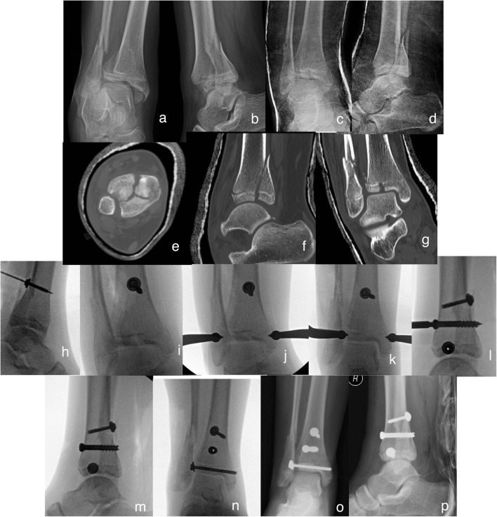
Fig. 6.
This triplane fracture in this 13-year-old female was sustained due to a fall (a, b). She was closed reduced under sedation and casted (c, d), then a CT scan obtained (e–g). Due to the proximal displacement of the posterior malleolar piece as noted on CT scan (f), decision was made to proceed with a posterolateral approach to the distal tibia and push fragment distally. An anti-glide screw was placed at the tip of the fracture line in anti-glide fashion (h). This was performed without drilling to allow the screw’s mass to push the fragment downward. Attention was then turned to the joint line without rigid fixation of the posterior piece to avoid over compressing the joint line and failing with reduction of the lateral joint fragment. Several projections were trialed to find this semi-external rotated view where the fracture line was noted to be most prominent (i). A large periarticular fracture clamp was then applied percutaneously and adequate reduction could be seen on fluoroscopy (j). The joint was fixed utilizing a 4.5 cannulated screw which lagged the fracture further (k, i). Finally, the posterior piece was fixed utilizing a 6.5 cannulated screw (l). Final radiographs reveal adequate joint line reduction (m–p)

