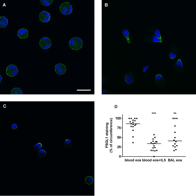
Fig. 6.
Localization of PSGL1 on blood and BAL eosinophils. Localization of P-selectin glycoprotein ligand-1 (PSGL1) (green) and nucleus (blue) in cytospun unactivated blood eosinophils (A), IL-5-activated blood eosinophils (IL-5, 50 ng/ml for 10 minutes as described in Materials and Methods) (B), and BAL eosinophils (C). Cells were analyzed by immunofluorescent staining using mAb to PSGL1 and AF488-conjugated secondary antibody. Nuclei were stained with DAPI. Bar, 10 µm. Representative of two experiments (i.e., 2 subjects, these 2 subjects were the same as used in Figure 5 but were different from the 6 subjects used in Figures 1–4). (D) Quantitation of PSGL1 localization using Fiji software, peripheral PSGL1 staining as percentage of circumference in unactivated blood eosinophils (blood eos), activated blood eosinophils (blood eos+IL5), and BAL eosinophils (BAL eos), **p < 0.01, ***p < 0.001 versus unactivated blood eosinophils.

