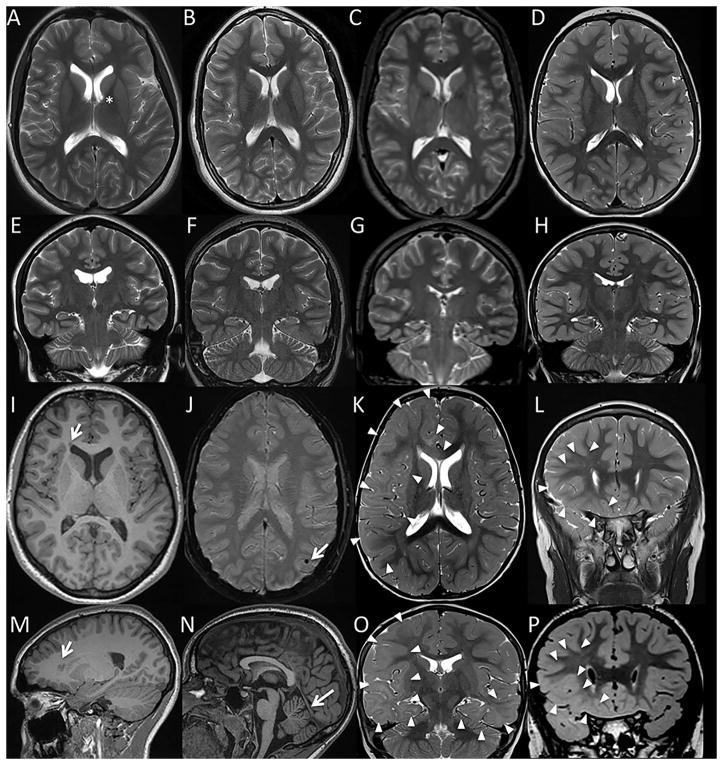
Figure 2.
Representative brain MR images from patients with somatic mutations in SLC35A2: Case 1 (A, E, I, M), Case 2 (B, F, J, N), Case 3 (C,G), Case 4 (K,O), and Case 5 (D, H, L, P). Axial T2 (A–D, K) and coronal T2 (E–H, L) images demonstrated grossly normal cerebral volume and preserved size of the hippocampi in all 5 subjects, allowing for some minimal prominence of the lateral ventricles and variant underrotation of the left hippocampus in Case 1 (E). For subjects Case 1, Case 2, and Case 3, there was no evidence of a malformation of cortical malformation apart from a tiny probable right frontal periventricular gray matter heterotopion in Case 1 (arrows in axial and sagittal T1 weighted images, I and M). For Cases 1, 2, and 3 there was also no evidence of a significant parenchymal injury or a metabolic/neurodegenerative condition, apart from nonspecific findings which were not thought to be related to seizure: an incidental left caudo-thalamic groove germinolytic cyst in Case 1 (asterisk in A); punctate mineralization versus hemosiderin deposition in the left temporo-parietal cortex of Case 2 (arrow in susceptibility weighted imaging, J); and some minimal cerebellar vermis volume loss in Case 3 (arrow sagittal T1 series, N). By comparison, subjects Case 4 and Case 5 demonstrated moderate to extensive parenchymal signal abnormality. In Case 4, T2 weighted imaging demonstrated diffuse haziness of the cerebral white matter concentrated in the right-greater-than-left frontotemporal lobes and right forceps minor (outlined by arrowheads in axial and coronal T2 weighted sequences, K and O). This pattern was consistent with cortical dysplasia and brain dysmyelination/gliosis; cortical dysplasia was thought unlikely to solely explain the findings given extent of signal abnormality, and tumor was thought unlikely given lack of mass effect. While T1 and T2 weighted imaging appeared grossly normal for subject Case 5, volumetric FLAIR imaging demonstrated loss of gray-white matter differentiation throughout the lower half of the right frontal lobe (arrowheads in coronal volumetric FLAIR, P; corresponding image slice on coronal T2, L). Although lacking a transmantle sign specific for cortical dysplasia, the regional blurring of gray-white matter differentiation in Case 5 was most suggestive of a focal cortical dysplasia; however, regional dysmyelination/gliosis could conceivably also have this appearance.

