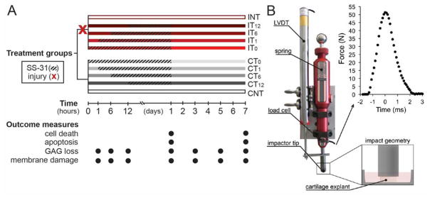
Figure 1. Experimental Methods. A) Study Design.
Half the cartilage explants were impacted (X) at time 0. Injured groups (I; red bars) and non-injured groups (C; grey bars) were then treated (hatched line) with SS-31 (1μM) at time zero (T0), 1 hour (T1), 6 hours (T6), or 12 hours (T12) after injury, or left untreated (NT). SS-31 treatment was withdrawn at 24 hours post-injury. Explants were imaged on day 1 or 7 for cell death or apoptosis, and cartilage conditioned medium was collected at 1, 6, and 12 hours, and 1, 3, 5, and 7 days after injury to assess glycosaminoglycan (GAG) loss and cell membrane damage.
B) Impactor setup and impact geometry. Modified from Bonnevie, et al., 2015. Reused with permission from SAGE Publications (pending).

