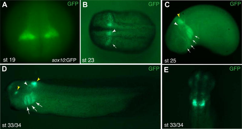
Figure 2. Spatial and temporal domains of the sox10:GFP reporter expression during development.

GFP reporter expression in Tg(sox10:GFP) embryos at different stages. A, B, stages 19, 23. White arrowhead in B marks hindbrain expression, the arrow points to migrating NC cells. C, stage 25. Major cranial migratory streams are indicated by arrows, white arrowhead points to the otic vesicle, yellow arrowhead to the expressing hindbrain domain. D, stage 33/34. GFP fluorescence is visible in the otic vesicle (white arrowhead), and three branchial arches (arrows). Yellow arrowheads point to the expression in the eye (a positive control GFP driven by the gamma-crystallin promoter, see Methods) and the hindbrain. (C, D) Lateral view is shown, anterior is to the left. (E) stage 33/34, dorsal view. Strong GFP signal remains in the hindbrain, weaker signals are detected in the forebrain and the otic vesicle.
