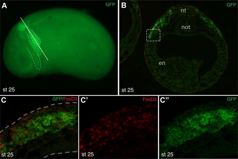
Figure 3. Characterization of GFP-positive cells in Tg(sox10:GFP) embryos.

A, Tg(sox10:GFP) embryo (stage 25) with GFP expression in the hindbrain and major migratory streams (dashed lines in A). Approximate position of the section in B is shown by a solid line. B, Cross-section at the hindbrain level of a transgenic embryo double immunostained for GFP and FoxD3. Nt, neural tube; not, notochord; en, endoderm. C, High magnification of the boxed area in B shows that the majority of GFP-expressing cells are FoxD3-positive. Dashed lines indicate tissue boundaries.
