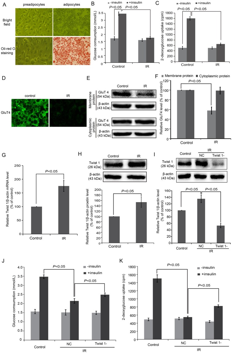
Figure 1.
An in vitro IR model was successfully established in cultured 3T3-L1 adipocytes. (A) Morphological observations and Oil red O staining of 3T3-L1 adipocytes. (B) Glucose consumption test showed low levels of glucose under insulin stimulation in IR cells. (C) Glucose uptake assay showed low levels of glucose uptake under insulin stimulation in IR cells. (D) Immunofluorescence detection revealed inhibition of GluT4 membrane translocation in IR cells. (E) GluT4 expression in the cell membranes decreased in IR cells according to western blot. (F) The semi-quantification of GluT4 (E) by western blot and using ImageJ software. (G) The mRNA of Twist 1 was determined in IR cells based on real-time PCR. (H) The expression of Twist 1 was determined in IR cells based on western blot. (I) The siRNA induced an obvious Twist 1 silencing effect according to western blot. (J) Glucose uptake tests revealed elevated glucose in Twist 1-silenced cells. (K) Glucose consumption assay also showed elevated glucose in Twist 1-silenced cells.

