Figure 4.
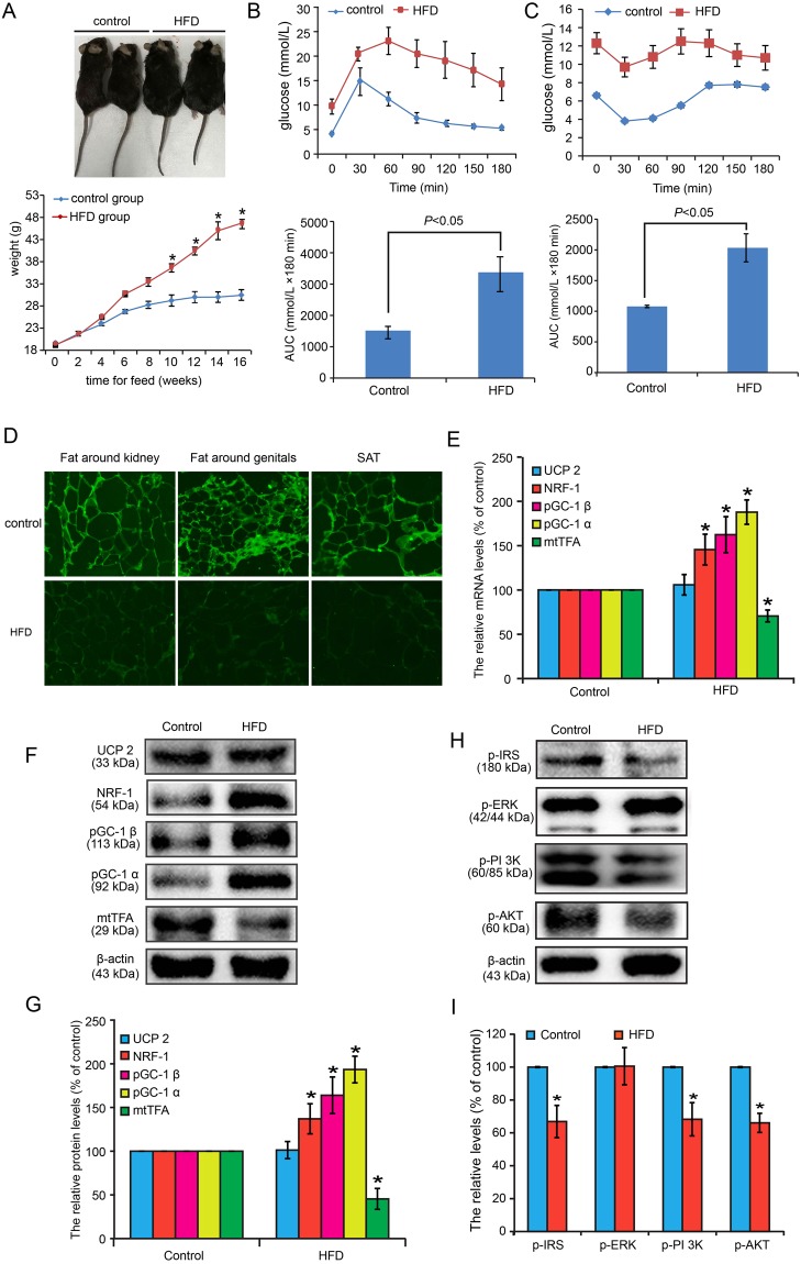
IR model in C57/BL6J mice was induced by continuous HFD feeding for 16 weeks. (A) Body shape and weight changes between control and IR groups. (B) IPGTT revealed that the sensitivity to glucose in IR was significantly decreased compared with controls. (C) IPITT revealed that the sensitivity to insulin in IR was significantly decreased compared with controls. (D) GluT4 membrane translocation in fat around the kidneys, fat around the genitals, and the SAT of IR mice were significantly decreased compared with control mice based on immunofluorescence by laser scanning confocal microscopy. (E) The mRNA levels of UCP2, NRF-1, pGC-1 α, pGC-1 β, and mtTFA were detected in mice perirenal adipose tissue. (F) The protein expressions of UCP 2, NRF-1, pGC-1 α, pGC-1 β, and mtTFA were monitored in mice perirenal adipose tissue. (G) The semi-quantification based on Image J software was conducted for (F). (H) Insulin signaling pathway in mice perirenal adipose tissue was also monitored after insulin (0.65 U/kg) stimulating for 10 min. (I) The semi-quantification based on Image J software was conducted for (H).

