Figure 5.
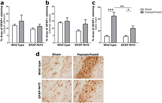
Astrogliosis in response to hypoperfusion was less severe in GFAP-Nrf2 animals. (a) % Area of GFAP+ staining in the corpus callosum and (b) internal capsule was unchanged following cerebral hypoperfusion. (c) % Area of GFAP+ staining in the optic tract was significantly increased by cerebral hypoperfusion (F(1,32) = 38.87, p < 0.0001), with a significant effect of genotype (F(1,32) = 7.45, p = 0.01). (d) Representative images of GFAP+ staining in the optic tract. Scale bar 100 µm. Mean ± SEM. Two-way ANOVA with Bonferroni adjustment for post hoc analysis. *p < 0.05, **p < 0.01, ***p < 0.001. n = 8–10 per group.

