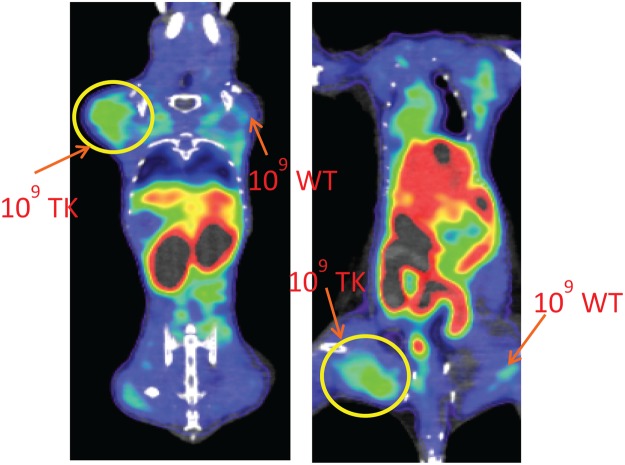
Figure 5.
In vivo PET images of mice infected with Pseudomonas aeruginosa (PAO1) and PAO1 tk-derivative (PAO1TK). Two BALB/c mice were i.m. infected with 109 CFU PAO1 (109 WT) or 109 CFU PAO1TK (109 TK) 2 h prior to i.v. injection with [18F]FIAU. WT (right upper and lower quadrants) infected mice and TK (left upper and lower quadrants) infected mice were subjected to PET/CT imaging 2 h post radiotracer injection. To obtain best views of the bacterial infection sites, the image planes showing upper and lower limbs of the animal were threshold adjusted to obtain the desired plane. Both the animals showed similar observations corresponding to injected bacteria. Data shown here are representative images from this experiment. Independent repeats of this experiment resulted in similar observation.

