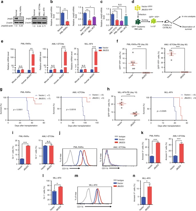
Fig. 2.
JMJD3 bears oncorepressor activity in mouse AML models. a, b Western blotting (a) or qRT-PCR (b) assay on Jmjd3 protein or mRNA level of GFP+ murine AML BM myeloid cell samples expressing PML-RARα, AML1-ETO9a, or MLL-AF9, and a normal sorted CD11b+ BM myeloid cell sample. c AML1-ETO, PML-RARα, or MLL-AF9 was transduced into normal c-Kit+ BM cells by retroviral infection, and the mRNA level of JMJD3 of the transduced GFP+ cells was measured by qRT-PCR. d Experimental protocol for testing the in vivo proliferative capacity of GFP+ BM leukemic cells transduced with YFP+ vector control or JMJD3-expressing vector in FVB/NJ or C57BL/6J mice. e GFP+ murine AML BM cells expressing PML-RARα, AML1-ETO9a, or MLL-AF9 were transduced with YFP+ empty vector or JMJD3-expressing vector, and the mRNA levels of mouse Jmjd3 or human JMJD3 were measured by qRT-PCR. f The percentages of PML-RARα (left panel) or AML1-ETO9a (right panel) GFP+YFP+ cells in the peripheral blood of syngeneic recipients after they had been transduced with YFP+ empty vector or JMJD3-expressing vector. g Kaplan–Meier curves are shown for syngeneic mice transplanted with GFP+ AML (left panel, PML-RARα+ cells; right panel, AML1-ETO9a+ cells) BM cell transduced with YFP+ empty vector or JMJD3-expressing vector. h The percentages of MLL-AF9+ GFP+YFP+ cells in the peripheral blood (left panel) and the survival (right panel) of the syngeneic recipients transduced with YFP+ empty vector or JMJD3-expressing vector. i, j Flow cytometric analyses of Gr-1 expression (i) or CD11b expression (j) on PML-RARα+ (left panel) or AML1-ETO9a+ (right panel) leukemia BM cells transduced with YFP+ empty vector or JMJD3-expressing vector and cultivated for 72 h. k Flow cytometric analyses of Annexin V staining on PML-RARα+ (left panel) or AML1-ETO9a+ (right panel) leukemia BM cells transduced with YFP+ empty vector or JMJD3-expressing vector. l–n Flow cytometric analyses of Gr-1 expression (l), CD11b expression (m), or Annexin V staining (n) on MLL-AF9+ BM cells transduced with YFP+ empty vector or JMJD3-expressing vector in vivo. Data are shown as the mean ± SEM; *p < 0.05, **p < 0.01, ***p < 0.001

