Figure 1.
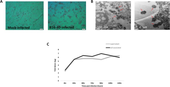
Virus culture and identification. (A) Mock-infected MARC 145 cells and bat paramyxovirus B16-40-infected MARC 145 cells showed cytopathic changes with light microscopy. (B) Bat paramyxovirus B16-40-infected MARC 145 cells imaged using transmission electron microscopy (TEM). (C) Viral growth kinetics for 6 days measured by infectious virus levels (TCID50/ml) in the supernatant and cell-associated sample. The time point of harvest was indicated as hours post infection.

