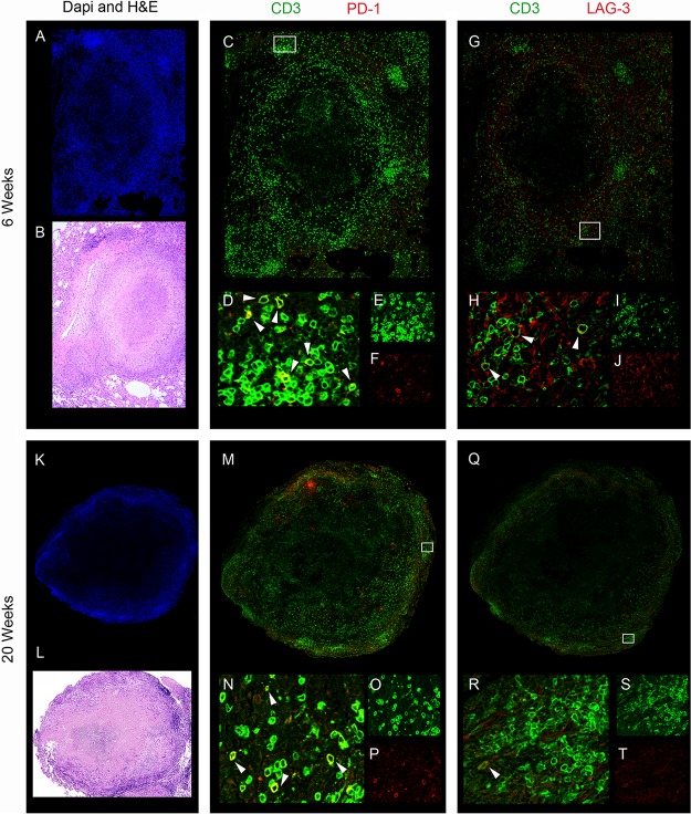
FIG 2.
Example of spatial organization of inhibitory receptors on T cells within TB granulomas. Few T cells in lung granulomas express PD-1 or LAG-3 inhibitory receptors, and most of the expression of these inhibitory receptors are on other CD3− cells. The rare CD3+ and PD-1+ or LAG-3+ coexpressing cells are localized in the periphery of the granuloma, away from the necrotic caseous center. Granulomas isolated at 6 weeks (A to J) and 20 weeks (K to T) were stained using antibodies against CD3, PD-1, and LAG-3 (see Materials and Methods for details). (A and K) DAPI staining of nuclei; (B and L) hematoxylin and eosin (H&E) staining; (C and M) merged image of CD3 (green) and PD-1 (red); (D and N) magnified region of interest (ROI) as indicated by a white box in panel C or M, where arrowheads indicate PD-1+ CD3+ cells; (E and O) magnified ROI showing green channel only (CD3); (F and P) magnified ROI showing red channel only (PD-1); (G and Q) merged image of CD3 (green) and LAG-3 (red); (H and R) magnified ROI, as indicated by a white box in panel G or Q, where arrowheads indicate LAG-3+ CD3+ cells; (I and S) magnified ROI showing green channel only (CD3); (J and T) magnified ROI of red channel only (LAG-3).

