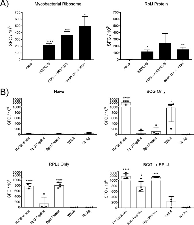
FIG 4.
BCG immunization does not impede ribosome-specific priming of CD4+ T cells. (A) Mice (C57BL/6) (n = 3) were immunized with 5 × 10⁷ CFU IKEPLUS i.v., 5 × 10⁶ CFU BCG s.c. followed by 5 × 10⁷ CFU IKEPLUS i.v. 4 weeks later, or 5 × 10⁷ CFU IKEPLUS i.v. followed by 5 × 10⁶ CFU BCG s.c. 2 weeks later. Two weeks after the final immunization, CD4+ T cells were purified from splenocytes and tested for IFN-γ production by an ELISPOT assay in response to ex vivo stimulation with recombinant RplJ protein (10 μg/ml) or purified mycobacterial ribosomes (5 nM). Responses that are significantly different from those in control naive mice by ANOVA are indicated (*, P < 0.05; **, P < 0.01; ***, P < 0.001; ****, P < 0.0001). Data shown represent mean values and standard errors for quadruplicate samples and are representative of results from three independent experiments. (B) Mice (C57BL/6) (n = 4) were immunized with 5 × 10⁶ CFU BCG s.c., 5 × 10⁶ CFU BCG s.c. followed 4 weeks later by two administrations of RplJ protein (25 μg) and CpG-ODN 1826 (20 μg) s.c. 2 weeks apart, or RplJ protein (25 μg) and CpG-ODN 1826 (20 μg) twice 2 weeks apart s.c. Two weeks after the final immunization, CD4+ T cells were purified from splenocytes and tested for IFN-γ production by an ELISPOT assay in response to ex vivo stimulation with recombinant RplJ protein (10 μg/ml), RplJTB146–160 peptide (10 μg/ml), or TB9.8 peptide (10 μg/ml). Positive-control wells were stimulated with the H37Rv sonicate (10 μg/ml). Responses that are significantly different from those of the no-antigen (No Ag) control by ANOVA are indicated (*, P < 0.05; **, P < 0.01; ***, P < 0.001; ****, P < 0.0001). Data shown represent mean values and standard errors for groups of four mice and are representative of results from two independent experiments.

