Fig. 1.
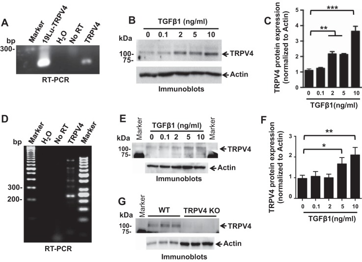
TRPV4 is expressed in primary normal human (HDFs) and mouse (MDFs) dermal fibroblasts, and stimulation of HDFs or MDFs with TGFβ1 produced a dose-dependent increase in total cellular TRPV4 protein expression. A: RT-PCR analysis demonstrating that TRPV4 mRNAs are present in HDFs. H2O (no RNA) and No RT (no reverse transcriptase) samples were used as negative control, whereas RNA extracted from human lung fibroblast cells (19Lu) was used as positive control. B: representative immunoblots show TRPV4 protein expression in HDFs treated with indicated doses of TGFβ1 for 48 h. Total actin was used as loading control. C: quantitative analysis of total cellular TRPV4 protein expression in TGFβ1-treated HDFs. Results shown are means ± SE from 3 independent experiments (**P < 0.01, ***P < 0.001 for TGFβ1-treated cells vs. untreated, n = 3, t-test). D: RT-PCR analysis demonstrating that TRPV4 mRNAs are present in MDFs. H2O (no RNA) and No RT (no reverse transcriptase) samples were used as negative control. E: representative immunoblots show TRPV4 protein expression in MDFs treated with indicated doses of TGFβ1 for 48 h. Total actin was used as loading control. F: quantitation of results from E. Results shown are means ± SE from 3 independent experiments (*P < 0.05, **P < 0.01 for TGFβ1-treated cells vs. untreated, n = 3, t-test). G: immunoblots show TRPV4 protein expression in wild-type (WT) MDFs, which is absent in TRPV4 KO MDFs. Triplicate samples were loaded for analysis and actin was used as loading control. For experiments described in this figure, cells were seeded on collagen-coated (10 μg/ml) plastic plates.

