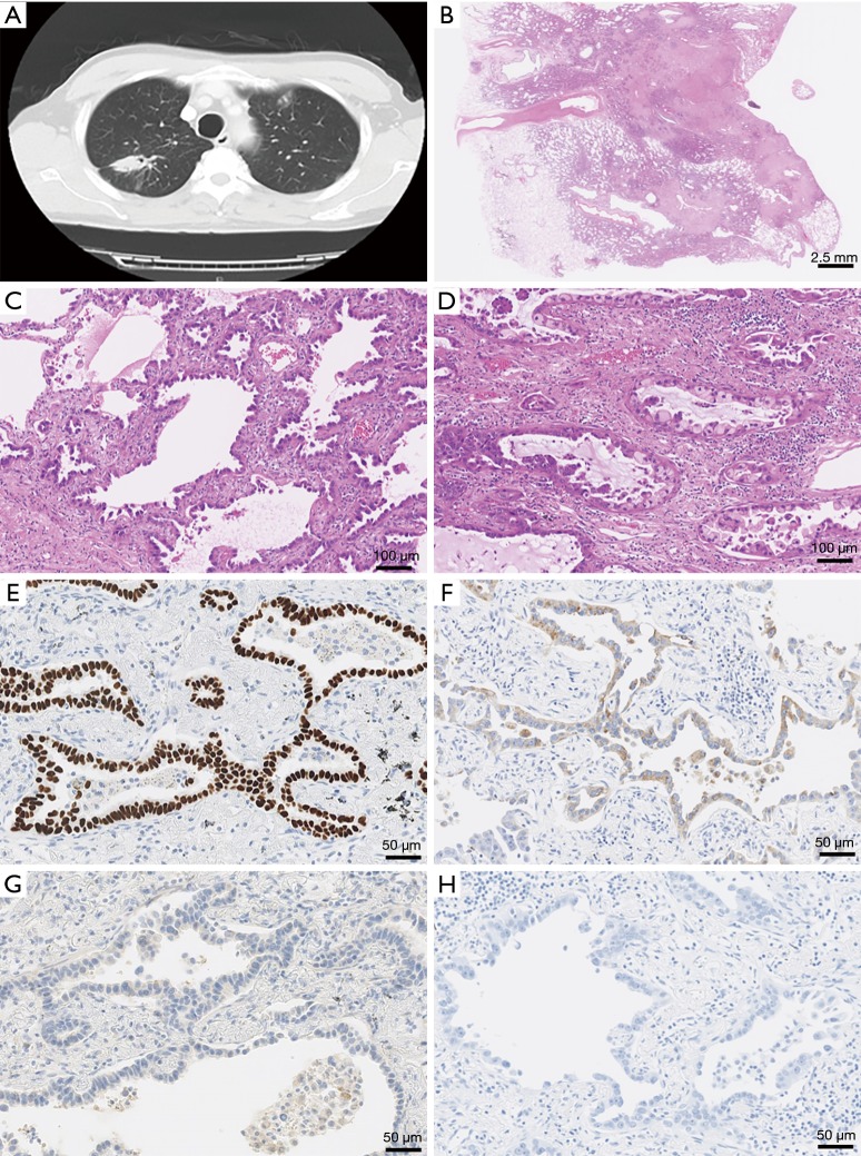
Figure 1.
Characteristics of primary lung carcinoma. (A) Chest computed tomography scan demonstrates a spiculated mass, measuring 33 mm × 17 mm in size in the right upper lobe; (B) a scanning magnification of it shows a proliferation of atypical epithelial cells with desmoplastic stroma [haematoxylin and eosin (HE) stain, magnification is ×1]; (C,D) histologic analysis shows a proliferation of atypical epithelial cells arranged in a papillary, lepidic or acinar growth structures (HE stains, ×100); (E,F) immunohistochemically, the specimen is positive for TTF-1 (E) and napsin A (F) (×200); (G,H) immunobiological staining of p40 (G) and CK5/6 (H) are negative (×200).

