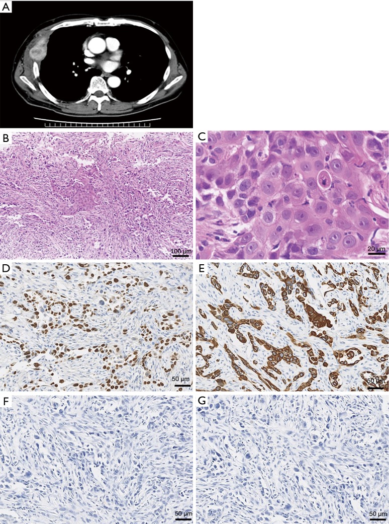
Figure 2.
Characteristics of pleural tumor. (A) Chest computed tomography scan demonstrates the pleural tumor destroying the right fifth rib; (B,C) histologic analysis shows a proliferation of atypical polygonal epithelial cells arranged in small nests or cords forming pseudoglandular structures with a few dyskeratotic cells and a desmoplastic stroma (B; HE stain ×100, C; HE stain ×200); (D,E) immunobiological staining of p40 (D) and CK5/6 (E) are positive (×200); (F,G) immunohistochemically, the specimen is negative for TTF-1 (F) or napsin A (G) (×200).

