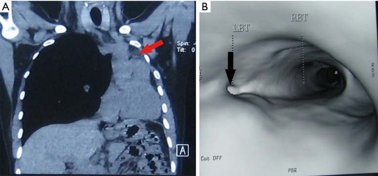
Figure 2.
The results of pre-operative examinations. (A) Chest CT image showing the left whole lung atelectasis (arrow) and a shift of mediastinum to the affected side, (B) bronchial three-dimensional reconstruction showing stenosis of the left main bronchus (arrow), and normal carinal structure was disappeared. LBT, left bronchus tunnel; RBT, right bronchus tunnel.

