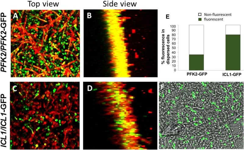
FIG 5 .
Carbon source utilization signature of biofilm and biofilm dispersal cells. C. albicans cells harboring a GFP-tagged glyoxylate pathway gene, ICL1 (ICL1/ICL1-GFP), and a GFP-tagged glycolytic gene (PFK2/PFK2-GFP) were allowed to form biofilms individually, for 48 h. The biofilm cells were stained with ConA, which stains the cell walls of the fungal cell red, and z-stacks were collected at two wavelengths, 488 nm (GFP) and 594 nm (ConA). (A to D) Top and side-scatter images reveal that the topmost hyphal layers of the biofilm expressed Pfk2 (A and B, respectively) rather than Icl1 (C and D, respectively). (E) In contrast, the cells dispersed from the biofilms expressed Icl1 at a significantly higher frequency than Pfk2. (F) Those results can be visualized by overlaying a bright-field image and GFP fluorescence image of the biofilm, with the result showing brightly fluorescent yeast cells on top of a mat of nonfluorescent ICL1-GFP biofilms.

