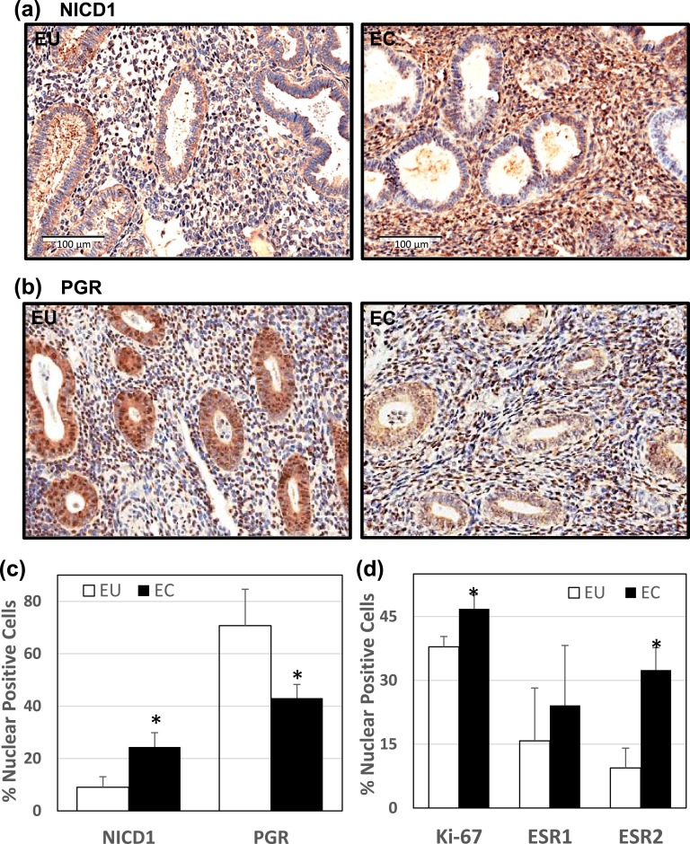
Figure 1.
Expression of NICD1 and PGR in EU vs ECs of women with peritoneal endometriosis. Formaldehyde-fixed, paraffin-embedded sections of paired EU and ECs from the same patient (UAMS cohort) were stained with anti-NICD1 or anti-PGR antibodies. Representative images of (a) NICD1 and (b) PGR immunostaining for EU vs ECs are shown. (c) The percentages of nuclear-localized, immunostained stromal cells for NICD1 and PGR were determined by counting the number of immunopositive nuclei over the total number of cells counted per field. Data (mean ± SD) represent analyses of tissue sections from n = 5 patients per group. For each tissue section, three to four random visual fields were counted. (d) Tissue sections were immunostained with anti-Ki67, anti-ESR1, and anti-ESR2 antibodies and analyzed as in c. Data (mean ± SD) are from n = 5 patients per group. For each tissue section, three to four random visual fields were counted. *P < 0.05 by Student paired t test between stromal EU and ECs.

