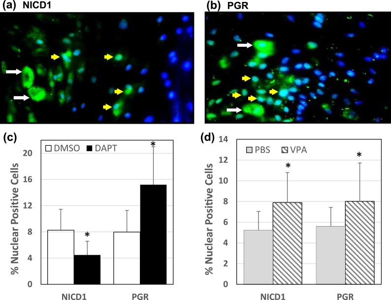
Figure 4.
Changes in nuclear-localized NICD1 and PGR in HESCs treated with DAPT or VPA. HESCs were treated with DAPT (10 μM), VPA (2 mM), or corresponding vehicle controls (DMSO for DAPT; PBS for VPA) and immunostained with anti-NICD1 and anti-PGR antibodies followed by incubation with fluorescence-conjugated secondary antibodies. Cells were counterstained with DAPI to identify nuclei. (a) Representative picture of anti-NICD1-immunostained and DAPI-counterstained cells after DAPT treatment. (b) Representative picture of anti-PGR-immunostained and DAPI-counterstained cells after VPA treatment. Nuclei were localized by DAPI (blue staining). For a and b, cytoplasmic-localized (green; white arrow) and nuclear-localized (blue-green; yellow arrow) proteins are shown. Nuclear-localized immunopositive cells for NICD1 and PGR were counted in cells treated with (c) DAPT or (d) VPA (and corresponding controls) and expressed relative to the total number of cells analyzed. Data (mean ± SD) are from two independent experiments, with three individual slides evaluated per treatment group per experiment. *P < 0.05 by Student t test between control and treatment groups for NICD1 and for PGR.

