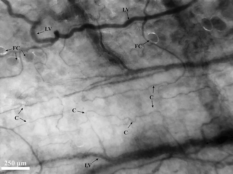
Fig. 2.
IDF image of the peritoneal microcirculation. A screenshot from a CytoCam video clip of the peritoneal microcirculation. Quadrangular network of large parallel vessels (LV) and their longitudinally orientated tortuous capillary (C) branches. The peritoneal microcirculation is often flanked by fat cells (FC).

