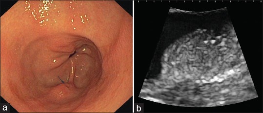
Figure 1.

Upper gastrointestinal endoscopy revealed a flat protruding subepithelial lesion in the gastric antrum (a) with central dimpling (arrow). Endoscopic ultrasound appearance (b); curvilinear echoendoscope UG EG-3870UTK, Pentax Medical, Hamburg, Germany with HI Vision Preirus (Hitachi Medical Systems, Wiesbaden, Germany) was consistent with ectopic pancreas: a slightly heterogeneous subepithelial lesion located within the submucosal layer. Histological proof of diagnosis was obtained after endoscopic resection
