
Figure 1.
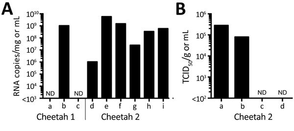
Detection of severe fever with thrombocytopenia syndrome virus (SFTSV) in samples from 2 cheetahs, Japan, 2017. A) RNA was extracted from tissues, plasma, and serum and subjected to quantitative reverse transcription PCR (RT-PCR). The amounts of SFTSV RNA were quantified, with a reference, as RNA copies/mg for tissues and RNA copies/mL for plasma and serum. The mean of duplicate results is shown in the graph. a, plasma; b, popliteal lymph node (left); c, serum; d, brain; e, salivary gland; f, spleen; g, mesentric lymph node; h, popliteal lymph node (left); i, popliteal lymph node (right). B) The TCID50 of salivary gland (per mg) and swab specimens (per mL) for cheetah 2 was determined using Huh-7 cells. Virus proteins were detected by an immunofluorescence assay with an anti-SFTSV N monoclonal antibody. a, salivary gland; b, oral swab sample; c, nasal swab sample; d, rectal swab sample. ND, not done; TCID50, 50% tissue culture infectious dose.