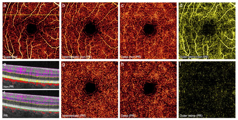
Figure 1. 64-year-old man without retinal pathology.
(A), 3×3mm optical coherence tomography angiogram (OCTA) of the superficial vascular complex (SVC). Vessels from the SVC project angiographic artifact onto en face images of the intermediate capillary plexus (ICP) (B), deep capillary plexus (DCP) (C), and outer retina (D). Projection-resolved (PR) OCTA removes artifact while maintaining vascular continuity of the ICP (G) and DCP (H), and accurately conveys the avascular nature of the outer retina (I). (E), Cross-sectional OCTA corresponding to the yellow line in (A) demonstrates retinal vessels (purple) with angiographic tails, and flow artifact in the outer retina (yellow). (F), PR-OCTA removes both the tail artifact and outer retinal flow.

