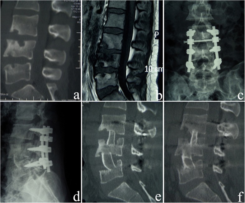
Fig. 1.

A 34-year-old female patient underwent anterior-posterior surgery and affected-vertebrae fixation. a The preoperative sagittal CT reconstruction image shows destruction of L3, L4, and L5 vertebrae and narrowing of the L3-L4 intervertebral space. b The preoperative sagittal contrast-enhanced MRI shows destruction of L3, L4, and L5 vertebrae and destruction of the L3-L4 and L4-L5 intervertebral disks. c, d One month after surgery, the anteroposterior and lateral X-ray images show that the fixation of the affected vertebrae is excellent. L3 and L4 vertebras are fixed with short pedicle screws. e Three months after surgery, the sagittal CT reconstruction shows that the lesion in the L3, L4, and L5 vertebrae has debrided completely, and the iliac bone grafts are firm. f Five years after surgery, the sagittal CT reconstruction shows that the L3–5 tuberculosis lesions are cured, and the bone graft fusion is solid
