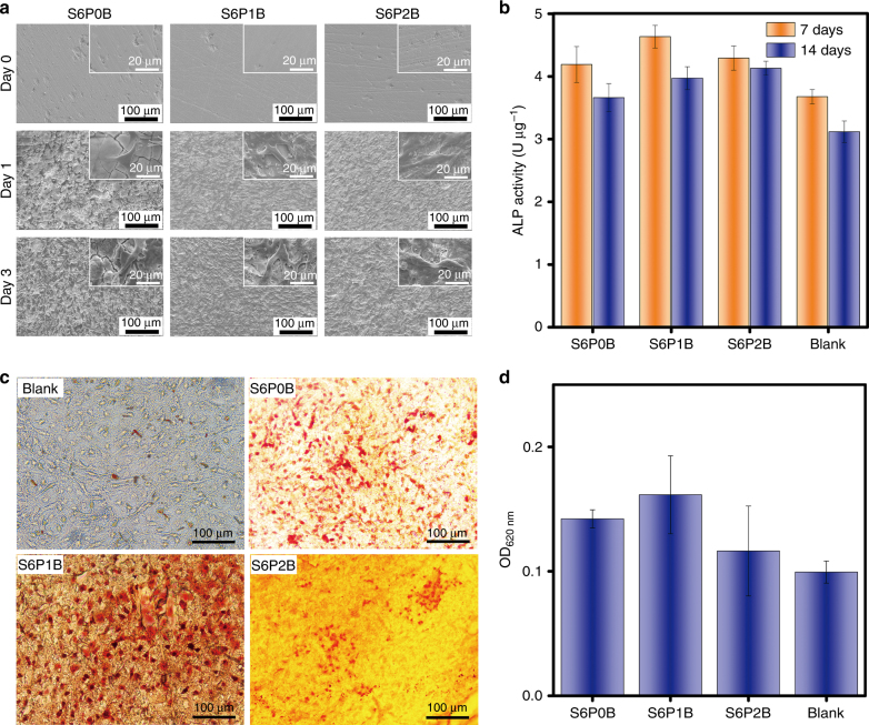
Fig. 6. Adhesion, proliferation, differentiation, and mineralization of murine pre-osteoblast cells on Bi-doped BGs.
a Adhesion and proliferation of MC3T3-E1 cells on the sample surfaces of S6PyB (y = 0, 1, 2 mol%) co-cultured for different days; b ALP activity of MC3T3-E1 cells cultured with S6PyB for 7 days and 14 days, respectively; c in vitro mineralization of osteoblast cells, reflected as calcium deposition on the surfaces of S6PyB, and blank sample co-cultured with MC3T3-E1 cells for 14 days and stained with Alizarin Red S; and d OD at 620 nm of S6PyB and blank sample co-cultured with MC3T3-E1 cells for 14 days, stained and dissolved by cetylpyridinium chloride. All data represent the mean values and error bars according to three independent experiments.

