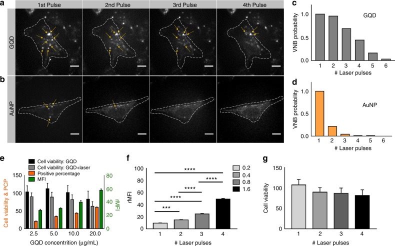
Fig. 1. Repeated VNB formation and photoporation of cells with GQDs.
HeLa cells were incubated with (a) GQDs and b AuNPs and irradiated with 4 discrete 7 ns laser pulses at twice the VNB generation threshold. Dark-field images show repeated formation of VNBs (yellow arrows) with each laser pulse for GQDs, but not for AuNPs. The cell boundaries are highlighted by the dashed lines. The scale bar is 20 µm. c The VNB formation probability from GQDs upon repeated laser exposure is calculated from the dark-field images (n = 25). d The same was done for AuNPs, showing that VNBs can only be formed once or, in rare cases, two times per AuNP. e Cell viability was measured by the MTT assay before (black bars) and after (gray bars) photoporation with GQDs (2.5 to 20 μg/mL) under the condition of a single-laser pulse. Cells were photoporated with FD10 to quantify the PCP (orange) and the amount of label delivered (green, rMFI relative mean fluorescence intensity). f HeLa cells were incubated with 10 µg/mL GQDs and photoporated four times with FD10. The concentration of FD10 was doubled at each step from 0.2 to 1.6 mg/mL to more clearly demonstrate successful subsequent intracellular delivery of FD10. **P < 0.01, ***P < 0.001, ****P < 0.0001. g Cell viability was measured after each laser treatment from 1 to 4 laser pulses. Error bars in (e, f), and g stand for three replicates

