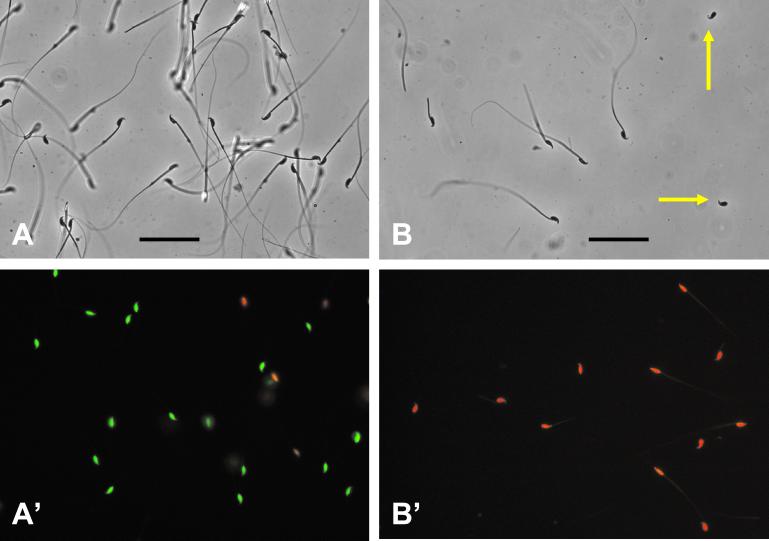
Figure 2.
Spermatozoa before and after freeze-drying. (A) Phase-contrast micrograph of spermatozoa in EGTA medium before freeze-drying, showing that most spermatozoa are structurally intact. (A′) Live/dead staining of spermatozoa in EGTA medium. Nuclei of “live” (plasma membrane-intact) spermatozoa fluoresce green, whereas those of “dead” (plasma membrane-disrupted) spermatozoa fluoresce red. (Bar equals 50 μm.) Spermatozoa in A and A′ are not in the same field of preparation. (B) Phase-contrast micrograph of freeze-dried spermatozoa, showing that many spermatozoa lost part or all of their tails (yellow arrows). (B′) Live/dead staining of freeze-dried spermatozoa, showing that all of them are “dead.”

