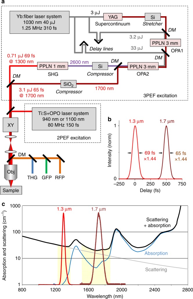
Fig. 1. Dual-band SWIR laser source optimized for three-photon microscopy.
a Experimental setup showing the source design. A Yb:fiber laser providing 1030 nm pulses at 1.25 MHz is used for supercontinuum generation in a YAG crystal and for amplification in a two-stage OPCPA arrangement. Signal and idler beams are produced at 1.7 and 2.6 µm and the idler is frequency-doubled, resulting in simultaneous emission at 1.7 and 1.3 µm with pulse energies in the µJ range. The beams are injected into a scanning microscope for three-photon microscopy. Alternatively, an 80 MHz pulse train at 920 or 1100 nm is used for comparison with two-photon excitation. DM dichroic mirrors, OPA optical parametric amplification stages, XY beam scanning, Obj microscope objective. b Measured temporal profiles for the two SWIR beams. c Parameters limiting deep-tissue imaging in multiphoton microscopy. The solid black curve combining tissue scattering and water absorption indicates the interest in use of the 1.3 and 1.7 µm wavelength ranges for in-depth imaging. The red and brown graphs reproduce the measured spectra for our source outputs, targeting the spectral regions of interest

