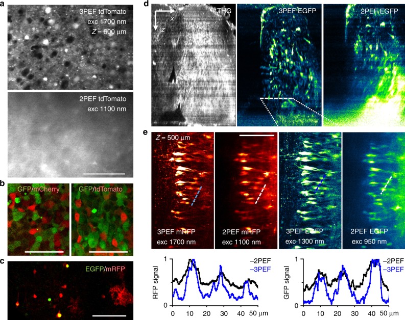
Fig. 2. Dual-color and in-depth 3P imaging of nervous tissues.
a Comparison of 3P and 2P excitation for imaging mouse brain tissue. 3PEF and 2PEF imaging of a tdTomato-labeled fixed mouse brain cortex at depths of 200 and 600 µm. See also Movie 1. b, c Dual-color 3PEF imaging for several combinations of green-red fluorescent proteins in b HEK cells and c mouse brain tissue at a depth of 500 µm. d, f Correlative 2PEF, 3PEF, and THG imaging of an intact chick embryo spinal cord (stage E9) co-labeled with EGFP and mRFP. Fluorescence images in each XY plane were normalized after acquisition for contrast comparison. See also Movies 2–4, and related information. d XZ projections of the THG, 3PEF EGFP, and 2PEF EGFP image stacks show the general morphology of the sample and the loss of 2PEF contrast with depth. e 3P and 2P mRFP and EGFP images recorded at a depth of 500 µm. Intensity profiles measured along the dashed lines illustrate the superior contrast provided in both channels by 3PEF excitation. Scale bars and arrows, 100 µm

