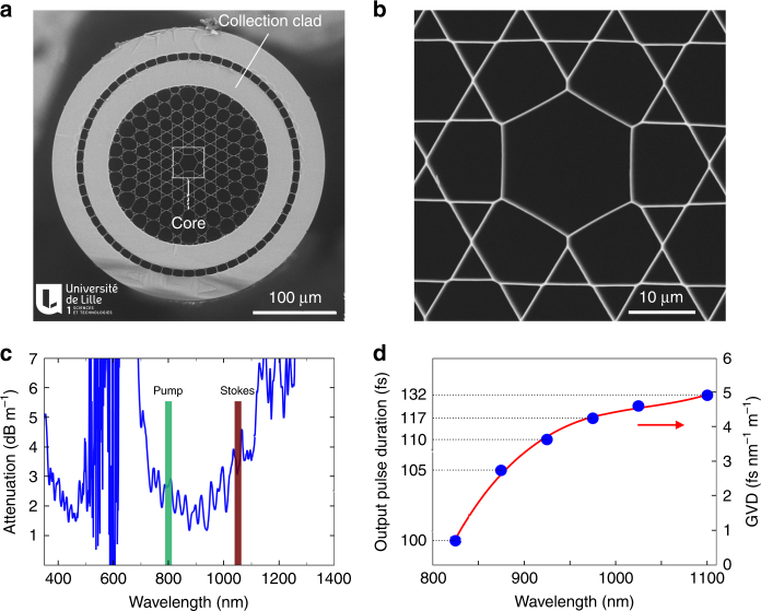
Fig. 2. Hollow-core photonic crystal fiber for multimodal nonlinear endoscopy.
a Electron microscope image of the hollow-core (HC) fiber featuring a Kagomé lattice together with a double cladding (DC) separated by air holes. The excitation pulses propagate through the HC, whereas the nonlinear signal is collected and transmitted through the DC. b Close-up view of the fiber core area. c Attenuation of the hollow fiber core; the wavelengths used for the pump and Stokes beams are highlighted. d Expected pulse duration after propagation over 1 m of fiber, for initially transform-limited 100 fs pulses. The output pulse duration is computed using the experimental GVD values (blue dots—right scale), which are obtained from the measurement of the group delay versus wavelength. The expected duration for a 150 fs input pulse centered at 1040 nm (the Stokes pulse) is 157 fs after 1 m of fiber. The red line is a third-order polynomial fit to the GVD data

