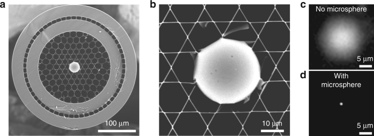
Fig. 3. Microsphere lens inserted into the hollow fiber core provides a submicron focus spot required for imaging.
a Scanning electron microscope image of the Kagomé HC fiber with a 30 µm silica microsphere inserted and sealed into the fiber core. b Close-up view of the microsphere inserted into the hollow core. c In the absence of the microsphere, the HC mode diameter is 15 µm, which is inappropriate for high-resolution nonlinear imaging. d The microsphere acts as a ball lens and focuses the light exiting the fiber core into a ~1 µm diameter spot

