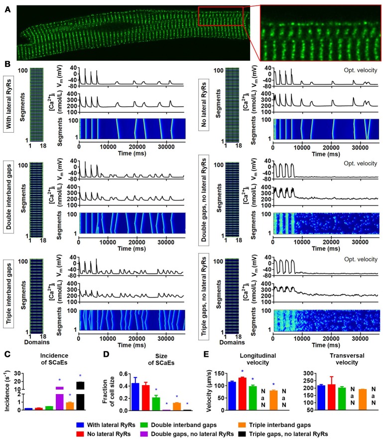
Figure 6.
Lateral RyR2s, inter-band distance and the propagation of SCaEs. (A) Confocal image showing an increased density of RyR2 close to the lateral membrane in a rabbit atrial cardiomyocyte. (B) Representative examples of membrane potential (VM), whole-cell Ca2+-transient, and longitudinal line scan for the 100-segment model with single (~2 μm; top row), double (middle row) or triple (bottom row) inter-band distance, with (left column) or without (right column) expression of lateral RyR2s between bands. In all panels, the total number of RyR2 was adjusted to achieve a density of 2,772 RyR2 per unit. The time constant of longitudinal Ca2+ diffusion between SR release spaces was adjusted from 0.22 to 0.07 ms in the model without lateral RyR2 expression to obtain similar SCaE properties for physiological inter-band distances. (C-E) SCaE incidence (C), size (D), as well as longitudinal and transversal velocity of Ca2+ waves (E) for the six model versions. An increased inter-band distance increased the incidence of SCaEs, reduced their size and altered the longitudinal velocity without affecting the transversal velocity. *indicates P < 0.05 vs. the control model (100-segment model of single inter-band distance with lateral RyRs; blue bars), n = 6 per condition.

