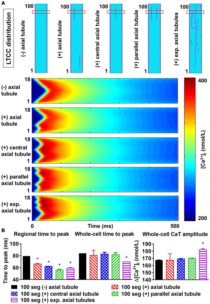
Figure 8.
Effects of axial tubule(s) on centripetal Ca2+-wave propagation. (A) Schematic representations of model structure: no axial tubule (L-type Ca2+-channels [LTCC] only at lateral membranes), single axial tubule at 33% of cell width, single central axial tubule at 50% of cell width, two parallel axial tubules at 33 and 66% of cell width, or experimentally observed axial tubules based on Brandenburg et al. (2016) (top) and corresponding transversal line scans of Ca2+-induced Ca2+ release during an action potential, showing centripetal Ca2+-wave propagation (bottom). (B) Quantification of time to peak (left) and Ca2+-transient amplitude (right) for the indicated line scan in the four groups (“regional”) or for the whole-cell Ca2+ transient. Increasing numbers of axial tubules and more centrally located axial tubules decrease the time-to-peak and slightly increase the Ca2+-transient amplitude. The magnitude of the increase depends on the number of axial tubules. *indicates P < 0.05 vs. the 100-segment group without axial tubules (black bars); n = 6 per condition.

