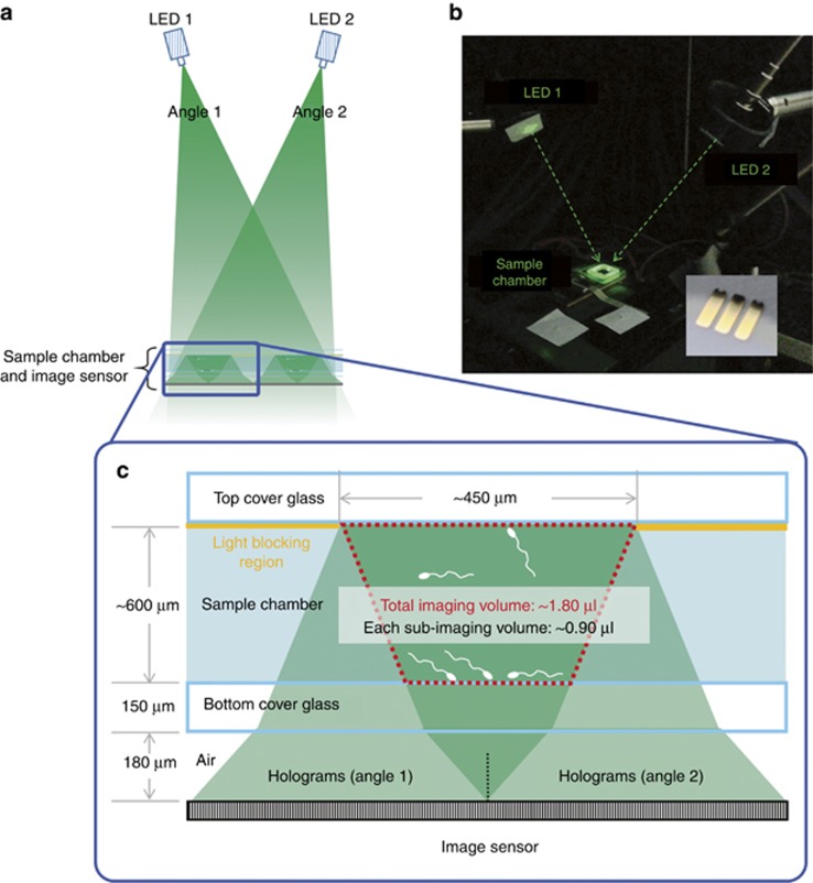
Figure 1.
Optical setup. (a) Dual-angle 3D sperm imaging and tracking platform using a spatially structured sample holder. (b) A photograph of the platform with the two fiber-coupled light-emitting diodes (LEDs, ~525-nm central wavelength with ~20-nm spectral bandwidth) placed at an angle of incidence of ~18° with mirror symmetry. The sample chamber is placed directly on top of the complementary metal oxide semiconductor (CMOS) image sensor, operating at ~300 fps. The inset is a photograph of the structured substrate that is generated by depositing gold (50-nm thick) on a glass slide. (c) Light passing through the mask generates a pair of spatially separated holograms for each sperm cell, fully utilizing the dynamic range of the image sensor and increasing the signal-to-noise ratio (SNR) of the reconstructions. The 3D imaging volume per bright stripe (space between the gold stripes) is 0.9 μl, resulting in a total imaging volume of 1.8 μl per experiment. The DOF is ~0.6 mm and the total volume of the sperm sample placed on the sample holder is ~34 μl.

