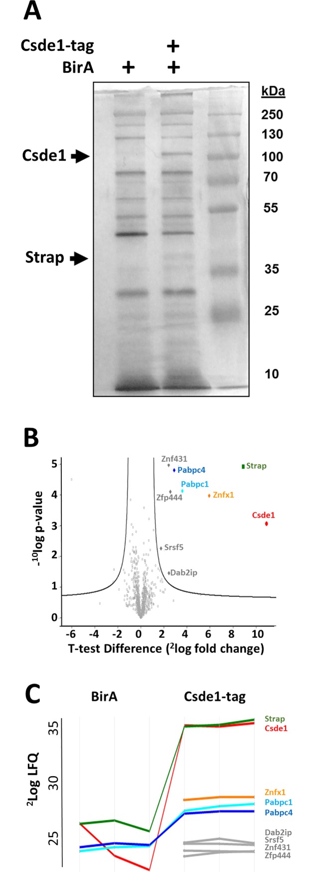
Fig 1. Proteins associated with Csde1 upon pull down of biotagged Csde1.

(A) Lysate of MEL cells expressing the biotin ligase BirA with or without biotagged Csde1 was incubated with streptavidin beads, washed, and eluted in laemmli buffer before loading on SDS-PAGE and silver staining. Third lane is a protein size ladder, sizes indicated in kD next to the lane. (B) Mass spectrometry of proteins pulled down by Csde1, and analysis by two-way t-test revealed 8 Csde1-associated proteins at a significance threshold of S0 = 0.8. -10log p-value is plotted against 2log fold change (n = 3). (C) Protein profile expression plot of Csde1-associated proteins (LFQ: Label free quantification). Lines are discontinued when peptides were not detected in control pull down in BirA only MEL cells.
