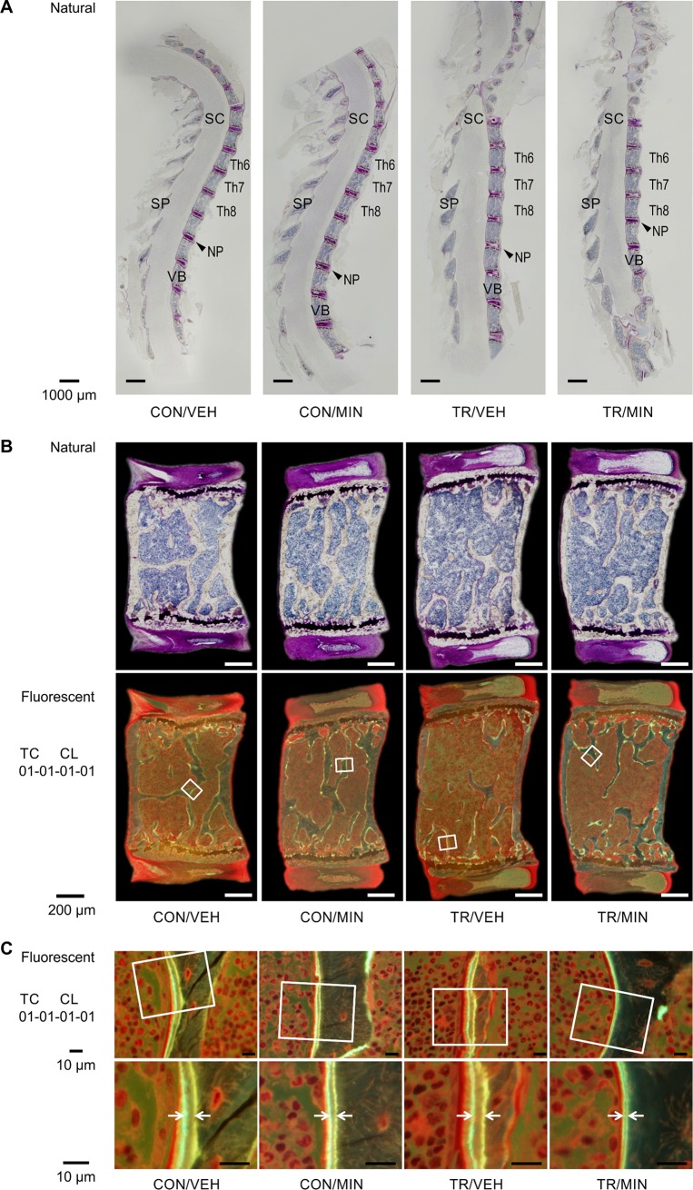
Fig 4. Bone histomorphometry in TR mice and effect of minodronate.
(A) Villanueva staining of undecalcified sagittal sections of the thoracic vertebral column of the CON/VEH, CON/MIN, TR/VEH, and TR/MIN mice. Bar = 1000μm. NP, nucleus pulposus; SC, spinal cord; SP, spinal process; VB, vertebral bodies. (B) Histological analysis of sagittal sections of the seventh thoracic vertebral column in each group. The trabecular bone volume in the TR/VEH group is lower than that in the CON/MIN group. The decrease was sufficiently inhibited by minodronate treatment. Trabecular bone, indicated by the white squares, is highlighted in Fig 4C. Scale Bar = 200μm. (C) Fluorescent images of representative sections of the vertebral trabecular surface demonstrating double tetracycline and calcein fluorochrome labeling. The enlarged figures show the increased distance between labeled bone surfaces in the TR mice compared with the untreated control mice. Minodronate preserves TR-induced increases in bone formation. Scale Bar = 10μm.

