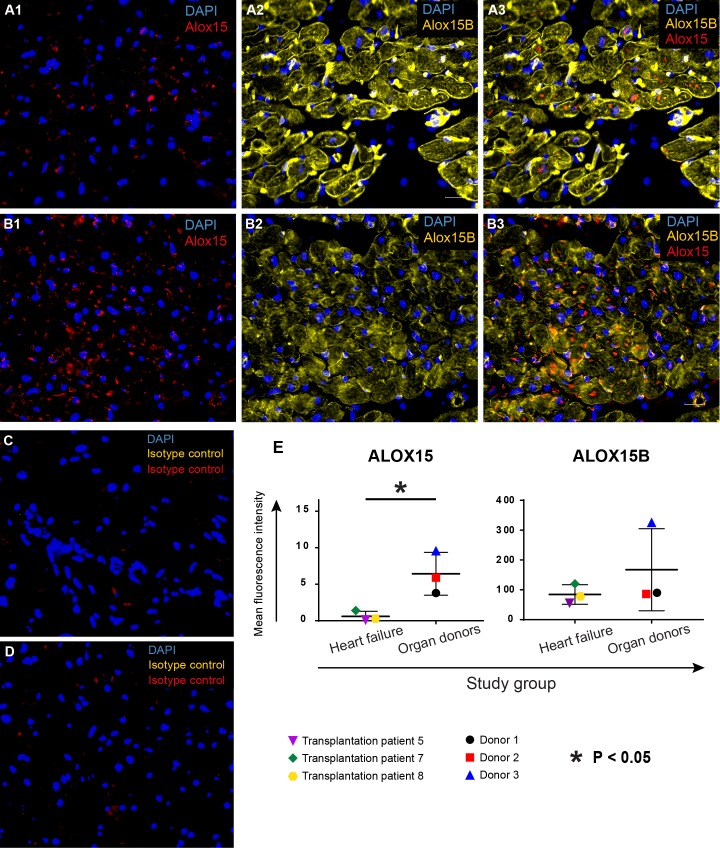
Fig 1. Expression of ALOX15 and ALOX15B in left ventricular tissue from failing human hearts and donor hearts.
Biopsies were snap frozen with liquid nitrogen and processed for IHC staining. Slides were incubated with primary antibodies for ALOX15 (red) and ALOX15B (yellow), followed by incubation with fluorochrome-conjugated secondary antibodes and mounting with DAPI (blue). (A) Representative images of ALOX15 and ALOX15B expression in the failing human heart. (B) Representative images of ALOX15 and ALOX15B in donor hearts not suffering from chronic heart failure. Corresponding isotype controls are shown for a failing heart (C) as well as for a donor heart (D). (E) Quantification of ALOX15 and ALOX15B expression. Both heart failure patients and donors expressed ALOX15 and ALOX15B, with a significantly higher expression of ALOX15 in donor hearts. Group wise statistical comparisons were performed using two-sided Student’s T-tests.

