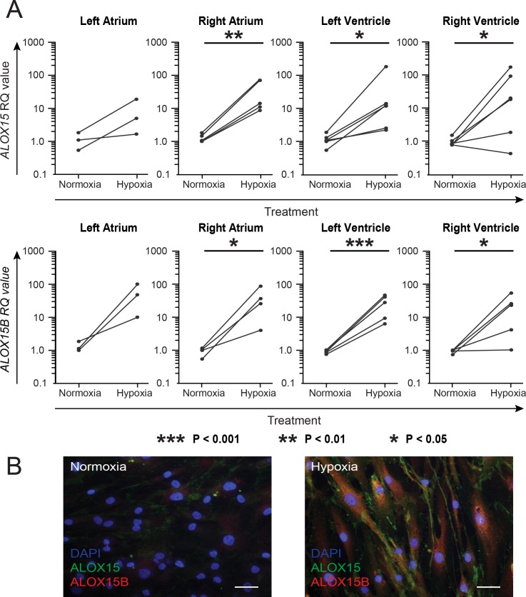
Fig 2. Expression of ALOX15 and ALOX15B in hypoxic human cardiac fibroblasts.
Cardiac fibroblasts were isolated from left (n = 3) and right (n = 5) atrium as well as left (n = 6) and right (n = 6) ventricle and subjected to 1% or 21% oxygen for 24 h. (A) Harvested cells were subjected to RT-qPCR and paired T-tests was performed using log-transformed RQ values. Paired RQ values are demonstrated as dots combined by a line. *** p < 0.001, **p < 0.01, *p < 0.05. (B) Immunocytochemical staining of human cardiac fibroblasts with primary antibodies against ALOX15 (green), ALOX15B (red) followed by flurochrome-conjugated secondary antibodies and DAPI (blue). Representative images of normoxic and hypoxic fibroblasts are shown. Scale bar = 40 μM.

