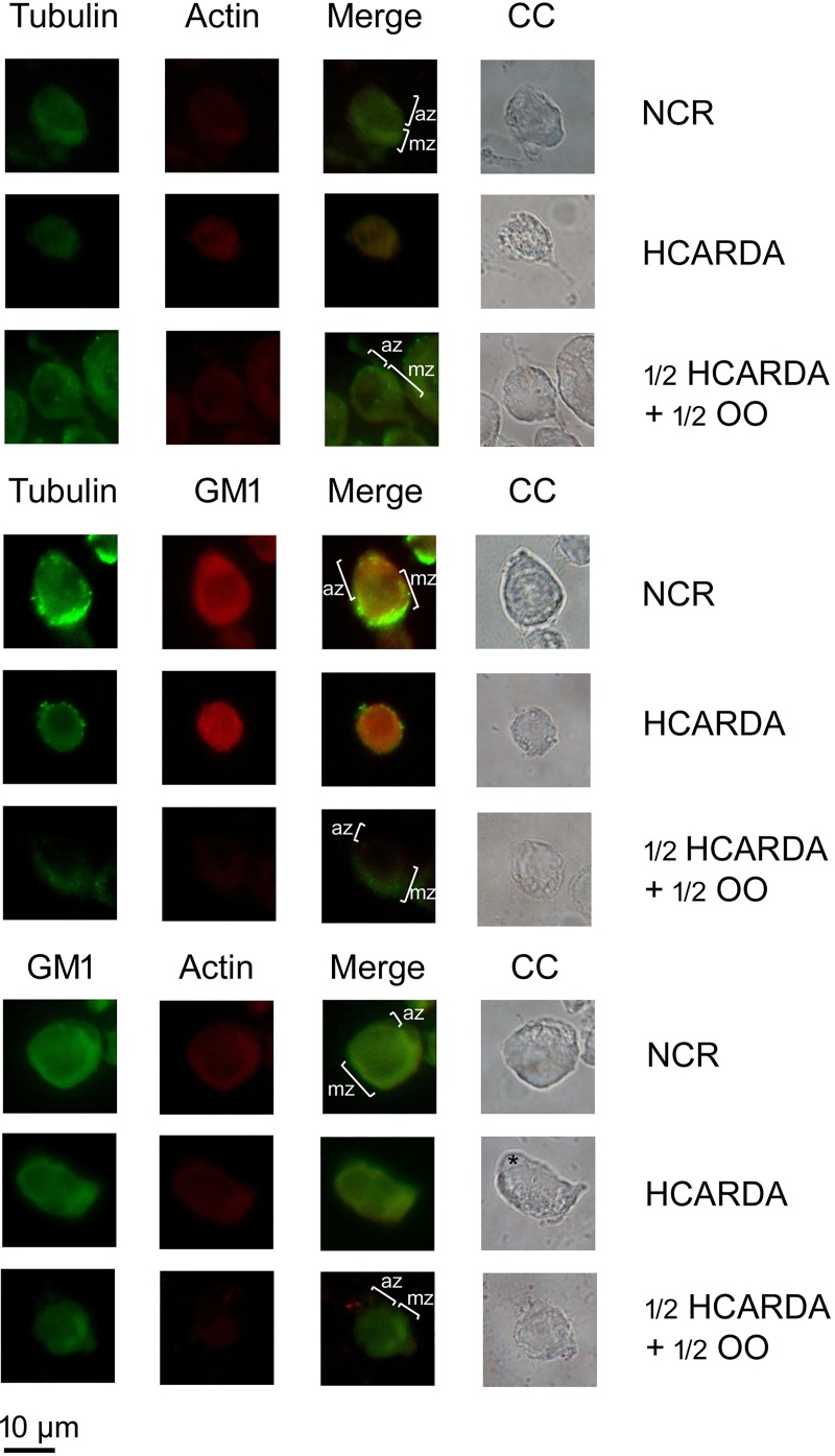
Fig 11. Microtubules, actin filaments and lipid rafts arrangement during spermiogenesis.
Spermatogenic isolated cells were analyzed to test the components of the sperm head elongation complex. The microtubules of the manchette were detected using alpha-tubulin antibody and secondary antibody combined with FITC (tubulin columns). The actin filaments were stained with actin antibody conjugated with Cy3 (actin columns). GM1-enriched lipid rafts were detected using cholera toxin conjugated with Alexa flour 594 (GM1 column, red signal) or FITC (GM1 column, green signal). The combination of green and red colors (merge columns) and phase contrast images were also included (CC columns). In NCR, microtubules, actin filaments and GM1 co-localized and were distributed in the manchette zone (mz) opposed to the acrosome zone (az). In HCARDA, it was not possible to detect manchette or acrosome zone. Microtubules, actin filaments and GM1 were equally distributed. Also, an asymmetrical acrosome was observed (asterisk). In ½ HCARDA + ½ OO, cells were polarized and manchette and acrosome zone were detected. Magnification: 620X.

