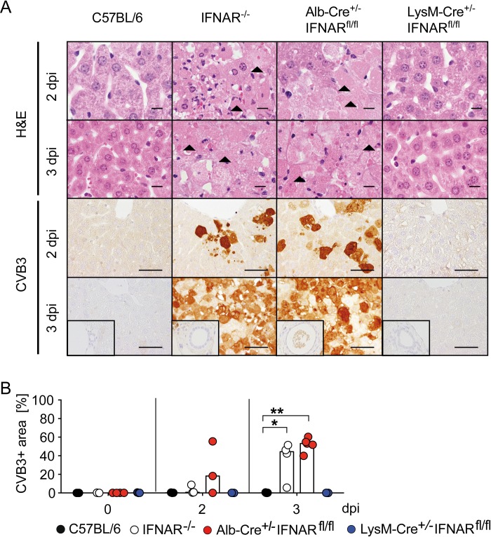
Fig 5. Mice with a hepatocyte-specific IFNAR ablation show infection of hepatocytes and severe hepatocellular necrosis.
C57BL/6, IFNAR-/-, Alb-Cre+/-IFNARfl/fl, and LysM-Cre+/-IFNARfl/fl mice were infected i.p. with 2 × 10⁴ PFU CVB3 and sacrificed 0, 2, or 3 dpi (n = 3–5). (A) Liver sections were H&E stained (line 1 and 2; bars = 10 μm) or subjected to CVB3-specific immunohistochemistry (line 3 and 4; bars = 50 μm). Arrowheads highlight necrotic hepatocytes (coagulative necrosis). Inserts show a lack of immunoreactivity in biliary ducts. (B) Quantification of area of infected liver tissue in CVB3-immunohistochemistry sections was performed by AnalySIS 3.2 software (n = 3–5). Bars depict median. Mann-Whitney test was used for statistical analysis, *P < 0.05; **P < 0.01.

