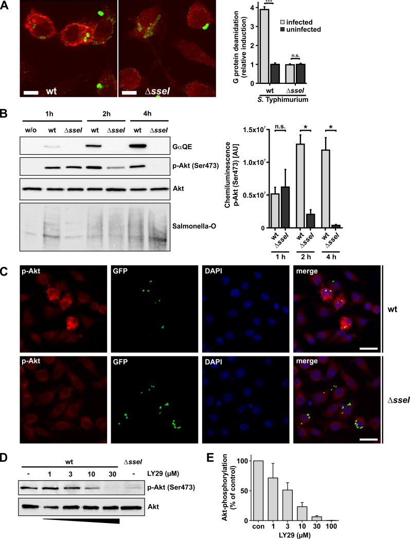
Fig 3. Cellular effects of SseI during infection.
(A) Fluorescence microscopy of fixed cells. RAW264.7 cells were infected with wild type (wt) S. Typhimurium or a ΔsseI-strain at a MOI of 1 for 5 h. Deamidation of Gα was detected utilizing the GαQE antibody and an Alexa 568-conjugated secondary antibody. Salmonella were identified by Salmonella O antiserum and an Alexa 488-conjugated secondary antibody. Deamidated Gα is depicted in red and Salmonella in green. Z planes showing Salmonella were maximum projected into one image. Scale bars = 5 μm. Quantification of images (right panel). Gα protein deamidation of Salmonella-infected or uninfected cells was calculated by determining the average intensity of Alexa 568 fluorescence of the whole cell area in the Z plane with internalized Salmonella. Significance was determined by two-tailed Student’s t-test. Data are means ±SEM. (n = 10 cells). (B-D) Serum-starved RAW264.7 cells were infected with a MOI of 30 for 30 min. (B) Time-resolved immunoblot analysis. At indicated times p.i., cells were lysed and processed for immunoblotting. Blotting membranes were treated with indicated antibodies (GαQE, p-Akt and Akt). Representative blots of 3 independent experiments are shown. Equal loading was verified by detection of Akt, presence of Salmonella was verified by Salmonella-O antigen staining. Right panel shows the quantification of p-Akt (Ser473) labeling over time after infection with wt- and ΔsseI-Salmonella strains from 3 independent experiments. Chemiluminescence intensity, given as area units [AU], was determined for each band. Statistical significance was assessed by Mann Whitney test. (C) Only macrophages infected with wild type (wt) Salmonella show strong phosphorylation of Akt. Cells were infected with pEGFP expressing wt- or ΔsseI-Salmonella Typhimurium for 30 min. 5 h p.i. cells were fixed and processed for immunofluorescence microscopy. Cells were stained for phospho-Akt Ser473 (red) and nuclei were stained with DAPI (blue). Scale bars = 20 μm. (D) Addition of the PI3K inhibitor LY29 leads to concentration dependent inhibition of Akt phosphorylation in wild type (wt) Salmonella-infected macrophages. 1.5 h p.i. cells were treated with the indicated concentrations of LY29 for 3.5 h. Thereafter, cells were lysed. Lysates were processed for immunoblotting with indicated antibodies (Akt, p-Akt(Ser473)). (E) Quantification of p-Akt (Ser473) immunoblots of infected cells treated with increased concentrations of LY29. Experiments were performed as depicted in (D). Intensity of labeled bands was normalized to untreated (con) cells. Values are means ±SEM from at least 3 independent experiments.

