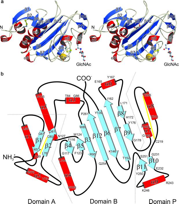
Figure 1.
Crystal structure of TL5A from the Japanese horseshoe crab T. tridentatus. (a) Stereo view of the TL5A ribbon plot. GlcNAc is represented by a ball-and-stick model and the Ca2+ ion is represented by a golden sphere. Disulfide bridges are colored yellow. Figure was prepared with molscript (28). (b) Topology diagram showing the arrangement of secondary-structure elements in TL5A. Disulfide bridges are indicated by yellow lines. Domains named in analogy to γ-fibrinogen fragment (21).

