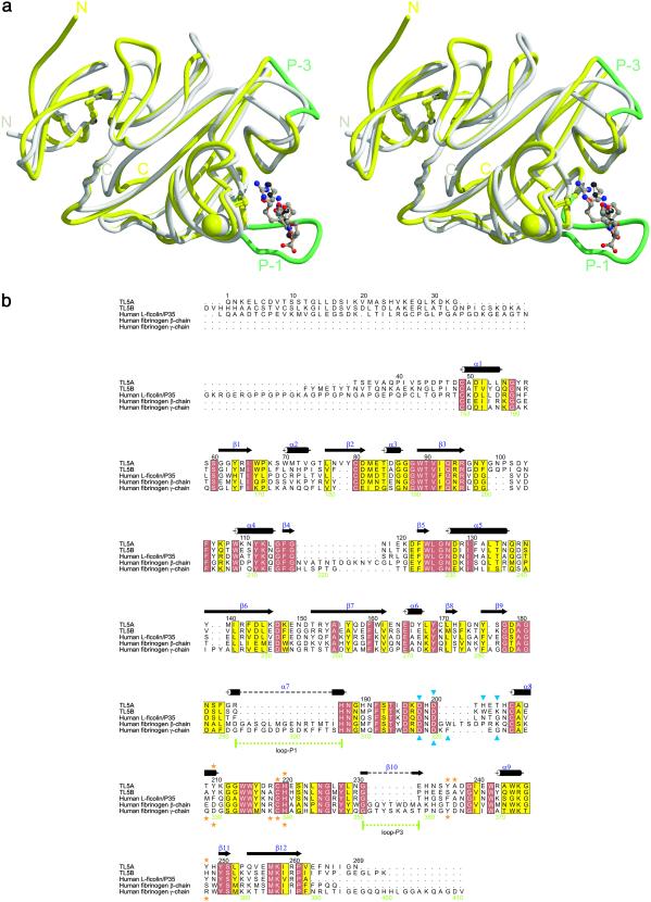
Figure 3.
Homology of TL5A and the γ chain fibrinogen fragment. (a) Superposition of the crystal structures of TL5A (gray) complexed with GlcNAc (white) and the γ chain fragment (yellow) complexed with GPRG-peptide (the A-knob, brown) (20). Ca2+ ions are represented by spheres of the same color as the structure they belong. (b) Sequence alignment of TL5A-related sequences. Black numbers on top refer to the TL5A sequence, and green numbers below refer to the fibrinogen γ chain sequence. Closed blue triangles indicate residues involved in Ca2+ binding. Orange stars indicate residues involved in GlcNAc (TL5A, above alignment), respectively A-knob binding (γ chain fragment, below). Figure was prepared with alscript (30).

