. 2018 Aug 9;7:e36654. doi: 10.7554/eLife.36654

Figure 6. VivosX of H2A.Z in human cells.

(A, B) Human MCF10A cells expressing an ectopic H2AFZ(H43C)V5 gene was incubated with 4-DPS for 20 min before lysis using the TUNES buffer. Total lysates were resolved by non-reducing SDS-PAGE in (A) and reducing SDS-PAGE in (B) before analyzed by anti-V5 immunoblotting. Z: nonubiquitylated H2A.Z; uZ: monoubiquitylated H2A.Z; ZxZ: Z-to-Z crosslink adducts; uZxZ: crosslink adducts with one uZ and one Z; uZxuZ: crosslink adducts of two uZ molecules.

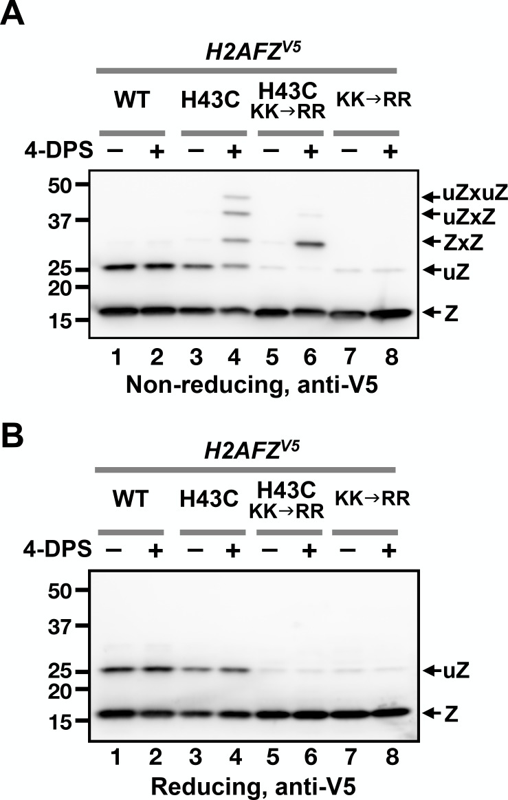
Figure 6.

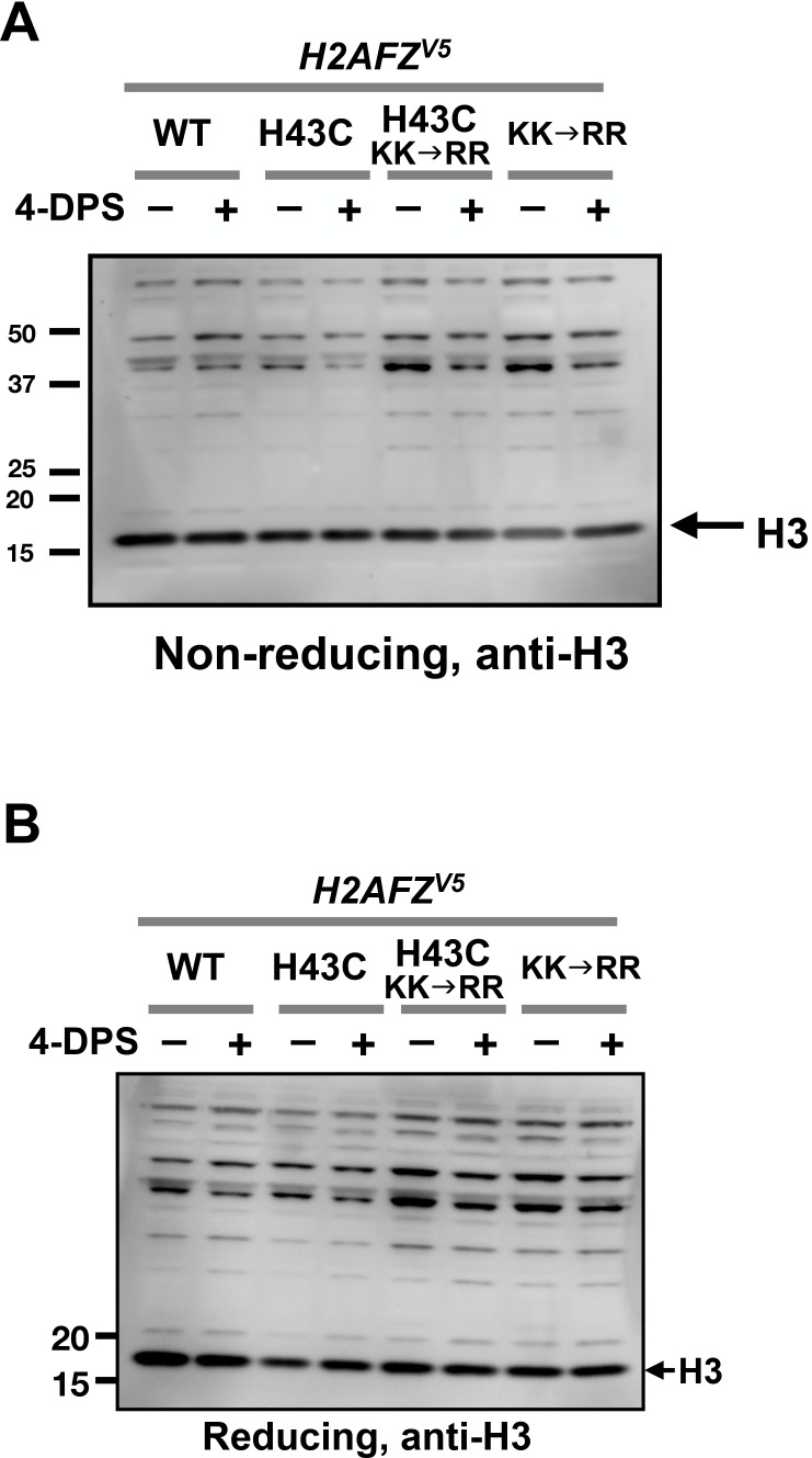
Figure 6—figure supplement 1. No apparent H3-to-H3 crosslinking observed after 4-DPS treatment.

Figure 6—figure supplement 1.

The protein extracts of MCF10A cells from Figure 6 were analyzed by non-reducing SDS-PAGE in (A) and reducing SDS-PAGE in (B) followed by anti-H3 immunoblotting.
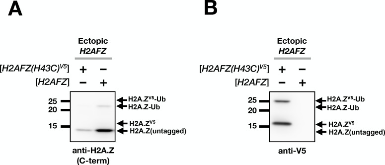
Figure 6—figure supplement 2. Estimation of ectopic H2A.Z level.

Figure 6—figure supplement 2.

(A) Total lysates of MCF10A cells transfected with H2AFZ(H46C)V5 or H2AFZ (untagged) driven by the same tetracycline-inducible promoter were analyzed by immunoblotting using an antibody directed against the C-terminus of human H2A.Z. (B) The same blot was probed with the anti-V5 antibody. Note that the H2A.Z antibody failed to detect the V5-tagged H2A.Z because the V5 tag at the C-terminus interfered with antibody recognition. In addition, the ubiquitylation sites of H2A.Z overlap the epitope of the anti-H2A.Z antibody. This may explain the disproportionately low H2A.Z-Ub signal in the anti-H2A.Z blot in comparison to the anti-V5 blot. The H2A.Z-Ub recognition bias by the C-terminus H2A.Z antibodies was previously observed (Sarcinella et al., 2007).