. 2018 Apr 25;42(4):338–342. doi: 10.4093/dmj.2017.0095

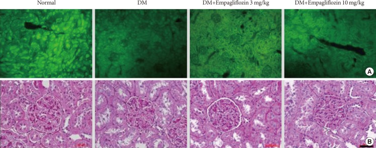
Fig. 2. Protein gene product (PGP) 9.5-positive small nerve fibers in the renal cortex and periodic acid-Schiff staining of kidney tissue sections. (A) Numerous PGP-9.5-stained small nerve fibers were observed in the empagliflozin-treated groups compared to the diabetes mellitus (DM) group. (B) The PAS-positive glomerular mesangial area was expanded in the DM group compared to the normal plus vehicle treated group. These changes were significantly ameliorated in the DM+E10 group; however, the effect was not significant in the DM+E3 group. Horizontal bar indicates 50 µm. DM+E3 and DM+E10, diabetes mellitus plus empagliflozin at 3 and 10 mg/kg, respectively.

Fig. 2