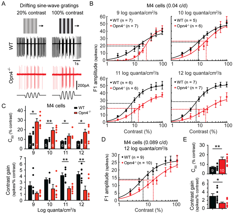
Figure 1. Melanopsin enhances the contrast sensitivity of M4 cells (ON alpha RGCs) across a wide range of physiological light levels.
(A) Example loose-patch recordings of WT (black) and Opn4−/− (red) M4 cell responses to drifting sine-wave gratings of 20% (left) or 100% (right) contrast in bright (12 log quanta/cm2/s) background light. Ex vivo retinas were presented with drifting sine-wave gratings of an empirically determined optimum spatial frequency (0.04 cycles/degree, Figure S1) of varying contrast. (B) Contrast response functions of M4 cells in WT (black) and Opn4−/− (red) retinas recorded at background light levels from 9 to 12 log quanta/cm2/s. Vertical dotted lines indicate C50 and horizontal dotted lines indicate half-maximal response. (C) C50 and contrast gain of WT (black) and Opn4−/− (red) M4 cells at background light levels from 9 to 12 log quanta/cm2/s. (D) Contrast response functions of M4 cells in WT (black) and Opn4−/− (red) retinas recorded in response to drifting sine-wave gratings with a spatial frequency of 0.089 cycles/degree. Recordings were made at bright background light levels (12 log quanta/cm2/s). (E) C50 and contrast gain of WT (black) and Opn4−/− (red) M4 cells in response to drifting gratings with a spatial frequency of 0.089 cycles/degree. All data are mean ± SEM. * P < 0.05. ** P < 0.01.

