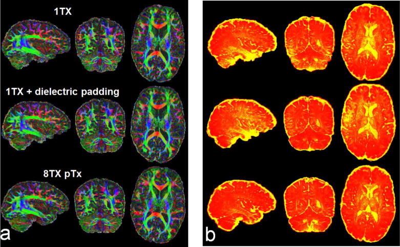
Fig. 3.

Comparing Nova single transmit (1Tx) coil alone (top row) and with dielectric padding (middle row), Nova 8Tx coil with pTx (bottom row) when acquiring high resolution whole brain dMRI at 7T with MB2. Shown are color fractional anisotropy (FA) maps (a) and corresponding sum of squared error (SSE) maps (b), derived by fitting a tensor model to the single-shell dMRI data acquired with inter-slice shift=FOV/2, in-plane acceleration factor=3 (iPAT3), TR/TE=7400/71 ms, acquisition time=10 min. For distortion correction, six b=0 images were acquired with same imaging parameters but inverted phase encode direction. The color FA is FA (in the range of [0 1]) with the color representing the orientation of the first eigenvector (red: left-right; green: anterior-posterior; blue: inferior-superior). The SSE map is shown in a colorscale of [0 12] (with yellow being high and red being low in SSE). Note that the use of pTx led to clear depiction of fibers not only in the cerebellum but also in the lower temporal lobe, and quantitatively, it reduced the whole brain average (excluding CSF) SSE by 22% relative to the combinational use of dielectric padding and Nova 1Tx coil, and by 24% relative to the use of Nova 1Tx coil alone.
