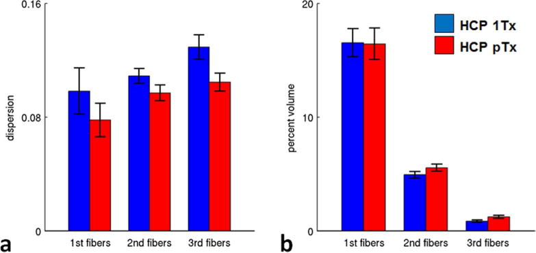
Fig. 6.

Fiber orientation estimation performance of HCP 7T dMRI protocol (HCP 1Tx) vs the HCP pTx protocol (HCP pTx) (five subjects). The dispersion (a) was significantly decreased for all of the three fiber orientation estimations (i.e., principal (p=3e-4, T(4)), second (p=1e-4, T(4)) and third (p=2e-4, T(4)) fiber orientations). The percent volume (b) was slightly decreased (p=0.11, T(4)) for principal fiber orientations, but increased significantly for both second (p=5e-4, T(4)) and third (p=1e-5, T(4)) fiber orientations when using pTx pulses. Error bars are standard deviation across the same five subjects.
